Konica KDR AU System Advanced U-Arm with DDR
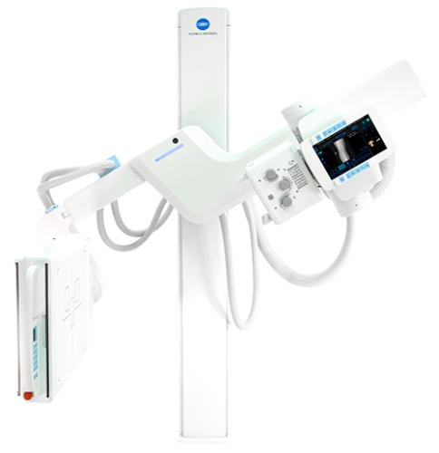
Konica KDR AU System Advanced U-Arm with DDR
The compact, efficient KDR Advanced U-Arm (AU) System with Dynamic Digital Radiography (DDR) features an array of advanced design innovations to optimize workflow, increase staff efficiency, outcomes, and now features Dynamic Digital Radiography (DDR).
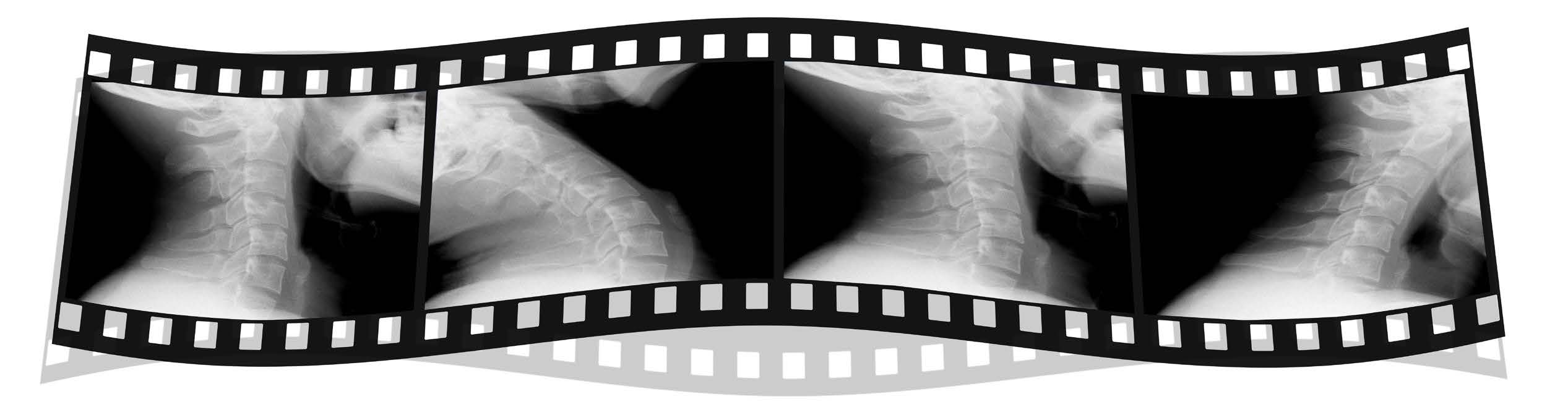
DDR is not Fluoroscopy, it is X-Ray that moves!
Product Description
Dynamic Digital Radiography (DDR) is a series of individual digital images acquired at high speed and low dose. The resulting cine loop presents a diagnostic-quality view of dynamic density and anatomical motion. Motion quantification is possible with proprietary advanced image processing.
Dynamic Digital Radiography supports the diagnosis of musculoskeletal conditions by presenting diagnostic detail in full motion. Fluoroscopy and cineradiography are proven diagnostic tools to examine joints and structures at rest and in motion to analyze biomechanics and musculoskeletal injury. The versatile, compact design of the KDR AU-DDR system makes it ideal for locations that need to provide superior-quality radiography services even in small rooms.
Konica Minolta's KDR-AU-DDR System employs a state-of-the-art, AeroDR HD 17x17 detector that maximizes efficiency, and delivers excellent bone and soft-tissue visualization from every study.
Specifications
| X-Ray system | |
| System Type | Floor Mounted U-Arm |
| Automation | Auto positioning pre-programmed U-Arm positions |
| Generator and Tube | Multiple configurations available |
| High Speed Motorized Movement | 1. Source to Image Distance (SID) 2. Arm Elevating Movement: Detector Arm/ Tube Arm 3. Rotation of Arm & Tube 4. Rotation of Bucky |
| Source to Image | 1000~1800mm (39.5~71 in.) |
| Vertical Travel | 40~170 cm (15.7~66.9 in.) |
| Arm Rotation Angle | -30°~120° |
| Tube Rotation | ±90° |
| Detector Rotation | ±45° |
| Motion Control | Tube head, detector and wireless remote control |
| Detector and Subsystems | |
| Built-in Detector | 17”x17” amorphous silicon digital detector |
| Scintillator (fluorescent substance) | CsI (Cesium iodide) |
| Detection Quantum Efficiency (DQE) | 65% @ 0.0 cyc/mm |
| Pixel Size | 175 um |
| A/D Conversion | 16 bit (65,536 gradients) |
| Dynamic Range | 4 digits |
| Image Area Size | 425.25 x 425.25 mm (2430 x 2430 pixels) |
| Preview Display Time | under 3 seconds |
| Exposure Interval (cycle time) | 3-5 seconds (wired connection) |
| Ultra Workstation | |
| System Platform | Desktop workstation with touchscreen – duplicate screen on tube head
DICOM 3.0 Compliant features – imaging, annotation, and analysis tools |